To streamline workflow and reduce cost of testing, researchers at Brown University and Columbia University have explored new single-cell analysis (SCA) technology and techniques in fresh and frozen tissues
Toward better cancer diagnostics, Brown University researchers have developed a new method of isolating single cells from complicated tissue biopsies. Single-cell analysis (SCA) has become an area of special focus because analyzing multiple cells at once does not allow for analysis of the genes, proteins, and other data that are specific to individual cells.
The Brown University project is important because of the difficulty in quickly pulling a single cell out of a mass of cells without damaging it. The complexity of this task creates a potential bottleneck in many forms of precision medicine testing, adding to the time, skill, and cost required to perform each test. To reduce some of the time-consuming steps involved, researchers sought to develop a technology to enable faster isolation of cells from biopsied cancer tissue to ready it for analysis, according to Anubhav Tripathi, PhD, study author and director of biomedical engineering at Brown University. The process uses electricity.
“Electric-Field Facilitated Rapid and Efficient Dissociation of Tissues Into Viable Single Cells,” from Scientific Reports in June, explains how freshly collected tissue from cells is placed in a fluid. An electrode is on either side. What happens when fluctuations occur in the electric field is that the cells should cleanly and completely separate from each other, without causing damage to them. The new process can isolate individual cells in as quickly as five minutes, according to the report.
Cell Sorting and Selective Cellular Recovery in Cancer Diagnostics
“Placing a tissue biopsy core within a liquid-filled cavity and applying an electric field between two parallel plate electrodes facilitates rapid dissociation of complex tissues into single cells,” the authors wrote, adding in a related Brown University press release that the process is unique.
The traditional approach and typical workflow would take a piece of a tumor removed in the operating room and brought to a lab, and use a process involving enzymes to extract nucleic acids from bulk tissue samples. Genetic sequencing would be performed en masse. The typical process results in low-resolution, potentially inaccurate genetic readouts, and poor detection of rare cell types, according to the study’s co-author, Nikos Tapinos, an associate professor of neurosurgery and neuroscience at Brown. The consequences of the loss of this information can be profound, including the possibility of patient misdiagnosis, creating a substantial lag between the time when the tumor is removed from the patient and the cells are ready for RNA sequencing.
Tapinos said there is an unmet need for technology that allows for the removal of tissue from the patient and, within minutes, the result of viable, healthy single cells from which RNA can be isolated.
New Technology at Brown Tests Cell Manipulation Using Electric Fields
“This [new] technology will be useful for those looking for answers using genomics, proteomics, transcriptomics; it will not only make those diagnostic and therapeutic investigations easier, but will also save researchers time and effort,” added Tripathi.
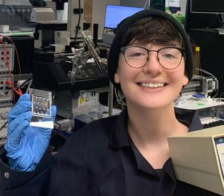
While Tapinos led the research, it was Cel Welch, a fourth-year PhD candidate at Brown, who is credited with inventing the new technology. “Dr. Tripathi has done a lot of work in his lab using electric fields and microfluidics,” Welch said.
“After seeing how electric fields could be used in other diagnostic applications, we had the idea to do something unique with the electric field that’s never been done before,” Welch added. “Based on the existing body of research about the manipulation of biological particles, we formulated a hypothesis about how this would work.”
SCA Technology Could Minimize Equipment Requirements, Enable Lower Processing Times and Costs
One of the key value propositions of this new technology is its simplicity when compared to existing workflows. “In the traditional workflow, you need to use several different lab instruments, such as a centrifugation instrument, that cost several thousands of dollars each,” Welch observed. “This approach to single-cell sample preparation requires only a single device.”
With the technology developed, the research team has applied for a patent and is focusing on developing a piece of equipment that can be easily used in a clinical setting. “A researcher will be able to simply press a button and, within minutes, have the single cells they need for analysis,” Tapinos said.
Columbia University Takes Up Single-Cell Analysis Using Frozen Samples
Single-cell analyses are leading to new advances in precision medicine on a regular basis. Another study published online in Cell by researchers at Columbia University, just two weeks after Brown’s announcement, relied entirely on single-cell analysis of frozen samples to discover ways that melanoma metastasis in the brain evades current treatment methods.
“Brain metastases are extremely common in patients with melanoma, but we have only had a rudimentary understanding of the underlying biology,” said Benjamin Izar, MD, PhD, assistant professor of medicine at Columbia University Vagelos College of Physicians and Surgeons, in a Columbia statement. “Our study gives us new insights into the genomics, immunology, and spatial organization of these tumors and serves as a foundation for further discovery and therapeutic exploration.”
The valuable insights of this study, however, were made more complicated by the difficulty of extracting single cells. “Recovery and interpretation of microglia single-cell transcriptomes from human tissues is challenging as these cells are particularly sensitive to fresh tissue processing and strongly express artifactual stress signatures,” researchers wrote, highlighting how difficult this process was.
While Columbia researchers were ultimately able to overcome the challenges of extracting single cells using more conventional methods, a tool like that developed by Tripathi’s team could have significantly simplified that step of the research.
With Brown researchers now focused on developing a tool for clinicians, healthcare leaders will recognize that this precision medicine testing resource may soon be available for clinical use. This advance has the potential to decrease costs and break bottlenecks in testing, improving the routine accessibility of precision medicine care.
—Caleb Williams
Related Information:
Electric-Field Facilitated Rapid and Efficient Dissociation of Tissues Into Viable Single Cells
Scientists at Brown Develop New Method and Device to Isolate Single Cells Using Electric Fields
Dissecting the Treatment-Naive Ecosystem of Human Melanoma Brain Metastasis
Columbia University Vagelos College of Physicians and Surgeons