Precision medicine treatments for glioblastomas have been highly researched due to the aggressiveness of this deadly cancer
Oncologists treating highly-aggressive glioblastomas may soon have a precision medicine tool that allows them to create an advanced fully-functioning model of a glioblastoma (GB) tumor. This model can inform the selection of a personalized medicine therapy tailored to a patient’s unique circumstances.
This is the kind of development that hospital and health system CEOs and administrators will want to follow because, as further research confirms the value of this approach, their institution could be an early-adopter of this technology. In turn, this would improve patient care and gain the hospital recognition for its state-of-the-art precision medicine oncology services.
The proof-of-concept study was conducted at the Tel Aviv University (TAU) Sackler School of Medicine in Israel. Researchers employed 3D bioprinting to create an advanced fully-functioning model of a glioblastoma (GB) tumor, a particularly dangerous and aggressive form of brain cancer.
The researchers believe their advanced GB cancer model will help oncologists determine which precision medicine treatments stand the best chance of succeeding within specific patients, according to a TAU news release.
In “Microengineered Perfusable 3D-Bioprinted Glioblastoma Model for In Vivo Mimicry of Tumor Microenvironment,” published in Science Advances, the researchers wrote, “We show that our model provides a more accurate evaluation of the native molecular and cellular characteristics of GB establishment and progression and the response to therapies compared to 2D plasticware models.
“Our 3D-bioprinted models can be exploited to better emulate the clinical scenario of various cancer types and can potentially serve as a powerful clinically accurate platform for preclinical research and drug discovery,” the researchers concluded.
In an article he penned for Johns Hopkins Health, Jon Weingart, MD, Director, Neurosurgical Operating Room at Johns Hopkins Medicine and Professor of Neurosurgery at Johns Hopkins University wrote, “If brain tumors were sharks, the glioblastoma multiforme, or GBM, would be the great white. More than any other brain cancer, GBM inspires fear because of its almost unstoppable aggression.”
Thus, oncologists and medical laboratories that treat such aggressive brain cancers may want to monitor the results of the Sackler School of Medicine study. It could provide significant advancements in precision medicine treatments for glioblastomas and other deadly cancers.
Mimicking In Vivo Conditions Ex Vivo
Standard treatment for glioblastomas includes a combination of surgery, radiation, and chemotherapy. Unfortunately, according to the National Brain Tumor Society, even with treatment, the average length of survival for a patient with GBM is only 12-18 months.
Because of the aggressive and dangerous nature of glioblastomas, this cancer has been an active area of research in precision medicine. The recent proof-of-concept study from the Sackler School of Medicine in Tel Aviv focused on providing lab-type cancer samples that can be experimented on ex vivo (out of the living) while still mimicking the conditions that actually occur in vivo inside each individual patient.
“Cancer, like all tissues, behaves very differently in a petri dish or test tube than it does in the human body,” said Ronit Satchi-Fainaro, PhD, Head of the Cancer Research and Nanomedicine Laboratory, and Hermann and Kurt Lion Chair in Nanosciences and Nanotechnologies, Department of Physiology and Pharmacology, Sackler Faculty of Medicine, at Tel Aviv University, in the TAU news release.
“Approximately 90% of all experimental drugs fail in clinical trials because the success achieved in the lab is not reproduced in patients. If we take a sample from a patient’s tumor, together with surrounding tissues, we can 3D-bioprint from this sample 100 tiny tumors and test many different drugs in various combinations to discover the optimal treatment for this specific tumor.” she added.
“Alternately, we can test numerous compounds on a 3D-bioprinted tumor and decide which is most promising for further development and investment as a potential drug,” she noted.
Recreating Fully Functional Cancer Models from Patients’ Own Tumor Cells
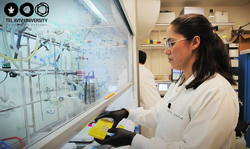
The Satchi-Fainaro Laboratory for Cancer Research and Nanomedicine developed a unique method for using 3D bioprinting to make fully-functional models of patients’ glioblastomas. “The process in which we bio-print a tumor from a patient is that we go to the operating suite, we take a chunk of the tumor, and we print it according to the MRI of that specific patient,” Satchi-Fainaro explained in a demonstration video of the technology.
“We have about two weeks in which we can test all the different therapies [that have] efficacy we would like to evaluate for that specific tumor and get back with an answer of which treatment is predicted to be the best fit,” she explained.

The 3D-printed tumor is printed in materials that mimic the conditions found in the brain. “The 3D-printed model is printed inside a bioreactor we have designed in our lab, and the print is made with a composition that mimics the brain,” said researcher and PhD candidate Lena Neufeld, who, according to the TAU new release, developed the 3D technology used to bioprint the cancer tumor. “We evaluate the cells and the composition to match the same tissues as we have in the human body.”
The researchers believe that a 3D representation of patients’ cancer—based their own cancer cells—provides an amazing opportunity. Clinicians can perform multiple treatments on each model, enabling them to experiment with treatment scenarios that would be impossible to do inside the patient themselves.
“The importance of our study is that this is the first time that we were able to 3D print a glioblastoma tumor, which is one of the most aggressive tumors in the brain,” explained Satchi-Fainaro in a video covering the 3D printing procedure.
Developing Novel Therapies for Precision Medicine Cancer Treatments
Satchi-Fainaro noted that the researchers also printed tiny blood vessels in the 3D printed tumors, which were connected to a pump that circulated the patient’s own blood through the tumor. “We pumped through them the blood of the same patient, but also monotherapy, one single therapy, or a combination of different treatments that are being used or being explored for glioblastomas.”
As a result of this approach, the 3D model better mimics the growth rate and behavior of each patient’s unique tumor, noted Eilam Yeini, PhD student and member of the TAU research team, in the video. “Through genetic sequencing, we showed that cancer cells that grow in our 3D models have a similar gene expression to the same cells growing in their natural environment, the brain,” he explained.
In addition to providing patient-specific treatment guidance, this research also could be used to safely develop and test new precision medicine treatment approaches.
“The impact of our research on future therapies for glioblastoma is that we will be able to use these 3D-printed models of glioblastoma to discover new targets for novel therapies that have, so far, never been used in treatments,” Satchi-Fainaro explained.
While the Satchi-Fainaro lab’s breakthrough represents a replicable breakthrough in treating glioblastomas using precision medicine, more research into the validity of 3D bioprinted cancer tumors must be completed.
Recent advances in providing precision medicine treatment for glioblastomas are an exciting beginning to this area of cancer research about which hospital CEOs and C-suite administrators should be aware. As new cancer research begins to result in practical clinical treatments, hospitals and health systems that are alert to these advanced therapies will be best positioned to implement them and reap the benefits they provide.
—Caleb Williams
Related Information:
Glioblastoma Multiforme (GBM)—Advancing Treatment for a Dangerous Brain Tumor
